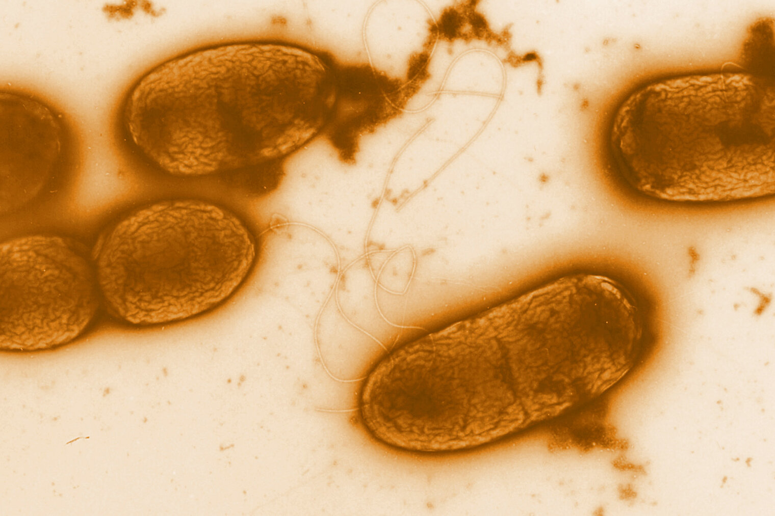
Electron micrograph
Electron micrograph of Pseudomonas aeruginosa
News

Discovery of molecular virulence mechanism of Pseudomonas aeruginosa

Structural biology insights by CSSB researchers reveal how pathogen breaks down host cell membranes and evades immune responses

Pseudomonas aeruginosa is a rod-shaped, Gram-negative bacterium capable of infecting various parts of the body, including the lungs, skin, and bloodstream. It accounts for approximately 7 per cent of hospital-acquired infections worldwide and is linked to around 559,000 deaths annually. Researchers from the group of Prof. Michael Kolbe at the Centre for Structural System Biology (CSSB), along with collaborators from Research Center Jülich, EMBL Hamburg, DESY and BIOTECH Thailand have uncovered novel virulence mechanisms of phospholipase C from P. aeruginosa. Their findings are published in Protein & Cell and shed new light on how this pathogen evades host defenses and causes severe disease. The CSSB is a common initiative by nine northern German research institutions, including the Helmholtz Centre for Infection Research (HZI) in Braunschweig.

Bacterial phospholipases (PLCs) are enzymes that breakdown lipids in cell membranes and are therefore essential virulence factors required for the efficient infection of host cells. Based on a study of clinical isolates conducted at HZI, Kolbe’s lab “Structural Infection Biology” identified PLC H and PLC N as relevant pathogenic factors in P. aeruginosa. “While we recognized that PLC H and PLC N played a significant role in infection, we had limited insight into how these enzymes actually function or what their structures looked like,” notes Michael Kolbe.

To elucidate the molecular mechanisms of PLC H and N, the researchers employed several structural biology methods to probe the biophysical and structural properties of the enzymes. Specifically, the team determined the structure of PLC H using cryo-electron microscopy (cryo-EM) at CSSB’s multi-user facility. Cryo-EM—a technique that captures high-resolution images of vitrified samples via high-energy electron beams— revealed that PLC H has three distinct molecular species.

To investigate how PLC N binds to different proteins, the researchers conducted X-ray crystallography experiments on the P11 beamline at PETRA III, DESY’s synchrotron radiation facility. “The combined cryo-EM and X-ray crystallography analyses showed that both enzymes have two domains with surprisingly similar structures,” explains Jörg Labahn, the study’s last author and a former CSSB group leader. 
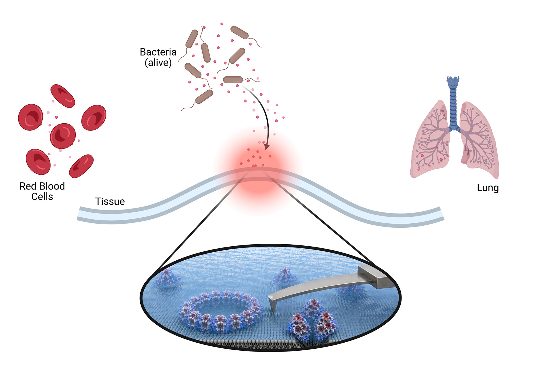
Structural Analysis of PLC H and PLC N
Bacterial phospholipases are potent virulence factors that hydrolyze ester bonds in membrane lipids and promote host cell damage during infection. Pseudomonas aeruginosa secretes two phospholipase C enzymes, PLC-N and the hemolytic PLC-H. Cryo-electron microscopy and X-ray crystallography revealed that both enzymes adopt a closely related tertiary architecture. Strikingly, time-resolved atomic force microscopy (AFM) showed that PLC-H rapidly perforates host membranes by generating large pores in lipid bilayers within minutes.

PLC H is the most abundant PLC found in P. aeruginosa and is most often found together with its chaperone R2. Notably, PLC H/R2 complex was found to have a much higher cytotoxicity i.e., a greater capacity to kill individual cells than PLC-N, despite their structural similarities. To investigate this phenomenon further, the researchers employed Atomic Force Microscopy (AFM), a high-resolution imaging technique that "feels" specimen surfaces using an atomically sharp probe. 

Using the AFM system available in the Barisch/Gutsmann Lab at CSSB, they performed time-resolved experiments revealing how PLC-H damages host cell membranes by forming large pores in lipid bilayers. "Remarkably," notes Udom Sae-Ueng, an AFM specialist who joined the project from BIOTEC, NSTDA, Thailand, "we observed pore formation in lipid bilayers within just seven minutes." Sae-Ueng’s involvement stems from a collaboration initiated after Her Royal Highness Princess Maha Chakri Sirindhorn of Thailand visited the DESY campus and CSSB in 2019.

“Investigating the phospholipases of Pseudomonas aeruginosa using complementary structural biology techniques, such as X-ray crystallography, small angle X-ray scattering (SAXS), cryo-EM and AFM, is only possible at CSSB,” explains Kolbe. "Thanks to its state-of-the-art in-house facilities and proximity to DESY’s PETRA III light source, this cutting-edge ecosystem has allowed us to uncover key molecular virulence mechanisms. These insights not only advance drug screening efforts but also hold potential for developing innovative therapies."

Original press release by CSSB
Original publication

Nishika Sabharwal, Yang Ge, Michele Lunelli, Udom Sae-Ueng, Cy M Jeffries, Spyros Chatziefthymiou, Sukrit Srivastava, Aziz Tumeh, Andre Geisler, Michael Kolbe, Jörg Labahn. Molecular virulence mechanism of phospholipase C from Pseudomonas aeruginosa, Protein & Cell, 2026;, pwag062, DOI: 10.1093/procel/pwag062

[Translate to English:] Charlotte Schwenner

Press contact

Dr Charlotte Schwenner
Science Editor