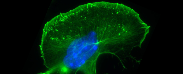
Cells in our body harbour a complex and frequently entangled network of filaments, which fulfils specific functions in maintaining or altering cell shapes during processes like embryonic development or migration of immune cells towards infectious agents. In addition, the cytoskeleton plays a central role in transport processes, both within cells and when crossing barriers between neighbouring cells. Therefore, it is not surprising that infectious agents have evolved numerous means to exploit the cytoskeleton of their hosts, which allows pathogens to enter host cells or to move across them to infect deeper tissue layers. “A better understanding of the cytoskeleton and the underlying molecular mechanisms of its formation will allow more precise interventions with pathogenic processes,” says Prof Klemens Rottner, head of the research group “Molecular Cell Biology” at the Helmholtz Centre for Infection Research (HZI) in Braunschweig.
In an international research project that has just appeared in the renowned scientific journal Science Advances, which was initiated by Prof Baoyu Chen from Iowa State University, USA, and substantially supported by the HZI-team around Klemens Rottner, the researchers were able to unearth novel, fundamental details concerning activation of the so-called WAVE Regulatory Complex (WRC). This protein complex assembles at the inner surface of the plasma membrane and operates like a central signalling unit. Upon activation, WRC triggers the formation of prominent cytoskeletal protrusions known as lamellipodia, which are key structures allowing the cell to migrate and engulf extracellular particles. As opposed to a static skeleton, the subpart of the cytoskeleton comprising actin filaments is highly dynamic: It is constantly re-arranged and thus enables cells to develop locomotive forces to drive cell migration and change cell shape.
It was previously shown that WRC is responsible for the assembly of actin filaments in lamellipodia downstream of the signalling switch Rac1 – a small GTPase. “That WRC activation was somehow connected to yet another GTPase, called Arf1, was also known,” Rottner says. “In the current study, we wanted to understand how precisely Arf1 docks onto WRC and what happens in the context of WRC activation upon Arf1 binding.”
Employing biochemical and structural biology approaches at Iowa State University and cell biological studies using CRISPR/Cas9 technology at HZI, together with diverse expertise from researchers at Stony Brook University, Mayo Clinic, and University of Pittsburgh, the team defined and characterized the Arf1 binding site on WRC and characterized its WRC-activating function.
“Specifically, WRC harbours three binding sites for signalling switches: two specific for Rac1 that were already established, and a novel one for Arf1. The latter is positioned between the two Rac-binding sites,” Rottner says. The researchers could also show that Arf1 can only bind WRC after Rac1 binding to its so-called D-site on the complex. “Rac1-binding to the D-site enables a conformational change on WRC, allowing interaction with Arf1. Such allosteric changes are famous phenomena in protein biochemistry, and in particular, in the context of WRC regulation,” Rottner explains. The researchers also established that optimal WRC activation occurs upon occupancy of all three GTPase binding sites. Together, the team was able to establish that aside from Rac1 binding, Arf1 constitutes a crucial signalling switch on WRC, required for its optimal function.
Such fundamental molecular insights as in this case on WRC regulation by Arf1 can be of potential interest for applied infection research. “Several previous studies have reported functions for Arf1 activation in host cell invasion by pathogenic bacteria – e.g., Salmonella. This was accompanied by increased actin remodelling enhancing by bacterial uptake,” Rottner says. “Arf1 or its binding site on WRC could thus constitute potential hubs for interfering with the pathogens’ invasion strategies.” Specific interference with this particular Arf1 mode of action might slow local actin remodelling and thus inhibit bacterial uptake. Rottner: “We hope that our insights will inspire the development of novel ideas and strategies for the intervention with specific host-pathogen interactions.”
Original publication:
Sheng Yang, Yubo Tang, Yijun Liu, Abbigale J. Brown, Matthias Schaks, Bojian Ding, Daniel A. Kramer, Magdalena Mietkowska, Li Ding, Olga Alekhina, Daniel D. Billadeau, Saikat Chowdhury, Junmei Wang, Klemens Rottner, Baoyu Chen: Arf GTPase activates the WAVE regulatory complex through a distinct binding site. Science Advances 2022 DOI: 10.1126/sciadv.add1412
Author: Nicole Silbermann


