The immune system is a highly efficient surveillance system that immediately takes action when, somewhere in the body, a cell signals that it is infected. A key mechanism of immune defence is based on cells carrying special receptors on their surfaces, which can display foreign protein fragments. If a foreign protein appears on the cell surface in the human leukocyte antigen (HLA) structures, special T cells (white blood cells) ensure that this cell is eliminated.
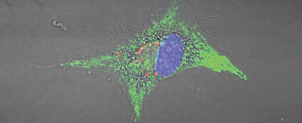
However, before the T cells can perform their monitoring duties in the body, they must be activated in the lymph nodes for specific antigens. Special immune cells perform this task, for example dendritic cells, which are able to present antigens on their surfaces. Dendritic cells are relatively rare in human bodies. That is why researchers did not know much about the uptake and processing of antigens and the presentation of antigen fragments on the human dendritic cells. Most of the findings to date were gained from experiments with mice and cell lineages.
Marius Döring, Hanna Blees and Nicole Koller, from the teams of Prof Ulrich Kalinke at TWINCORE and Prof Robert Tampé from the Institute of Biochemistry at the Goethe University Frankfurt, collaborated closely for several years in order to better understand how and when human dendritic cells display their antigens. “This mechanism represents the basis for the development of new and improved vaccines,” says Ulrich Kalinke from TWINCORE. “When working with human dendritic cells, many standard methods fail because these cells cannot be isolated
in sufficient numbers. Therefore we had to develope some further methods.”
The researchers asked themselves how it was possible that pathogen components are taken up at the site of infection, but that the critical protein fragments are only later presented to the T cells in the lymph nodes until later. Experiments with mice cells have shown that the loading of HLA receptor structures with protein fragments depends on a transport molecule called TAP (transporter associated with antigen processing). The scientists found that during the development of progenitor cells in dendritic immune cells, the number of TAP molecules increases, whereas the transport function decreases. “The reduced function of the TAP transporter also has a positive effect on the interaction between the dendritic cells and the T cells,” says Nicole Koller, scientist at TWINCORE and the Goethe University. Interestingly, the completed dendritic cells display antigens much better than progenitor cells and are thus more effective T cell activators.
“Furthermore, we were also able to resolve the contradictory finding that more TAP molecules with lower activity were found. The cause is a redistribution of TAP in the cell during the differentiation of the dendritic cells,” says Marius Döring, TWINCORE scientist in Ulrich Kalinke's team and now at the Institut Curie in Paris. In the progenitor cells, TAP was mainly found in the endosomal compartment, but was later transferred to the endoplasmic reticulum and the lysosomes during the differentiation to dendritic cells. “In this way, the absorption of foreign components, their fragmentation and the presentation of these fragments is separated, in terms of time and space, through the reprogramming of the progenitor cells into dendritic cells,” adds Hanna Blees, scientist in Robert Tampé's team and now at the Francis Crick Institute in London.
This research is certainly just the first step towards a better understanding of the function of human dendritic cells, states Ulrich Kalinke. “Nevertheless, this study
is an important breakthrough in developing improved vaccines.”
Original publication:
Marius Döring, Hanna Blees, Nicole Koller, Sabine Tischer-Zimmermann, Mathias Müsken, Frederik Henrich, Jennifer Becker, Elena Grabski, Junxi Wang, Hans Janssen, Werner Zuschratter, Jacques Neefjes, Frank Klawonn, Britta Eiz-Vesper, Robert Tampé, Ulrich Kalinke: Modulation of TAP-dependent antigen compartmentalization during human monocyte-to-DC differentiation. Blood Advances 2019; doi: 10.1182/bloodadvances.2018027268


